
Glossary
Constitutions
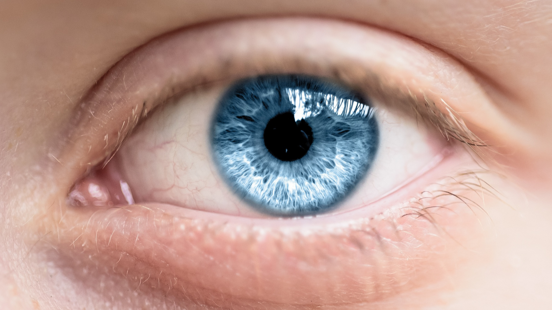
Lymphatic
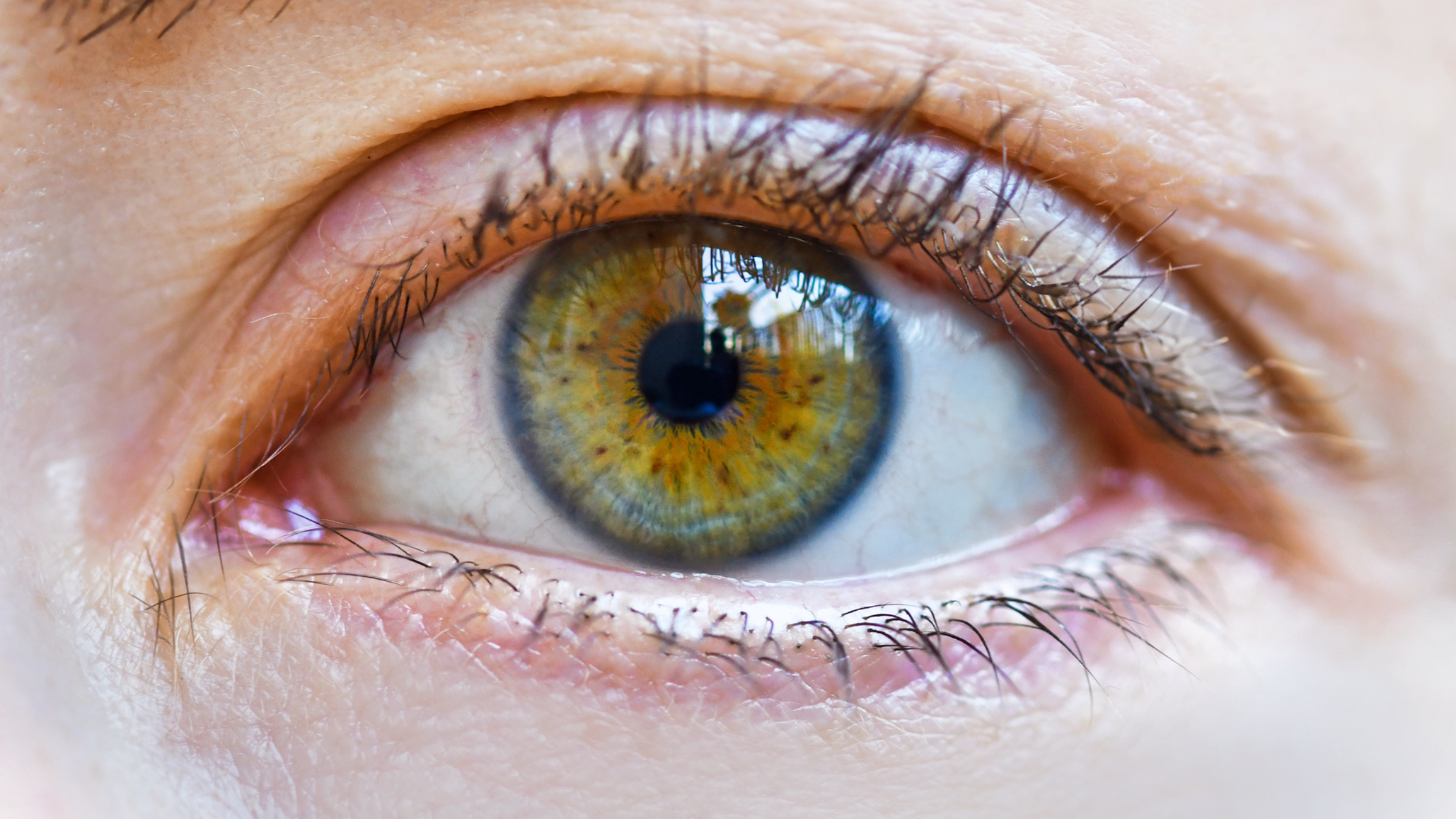
Mixed Biliary
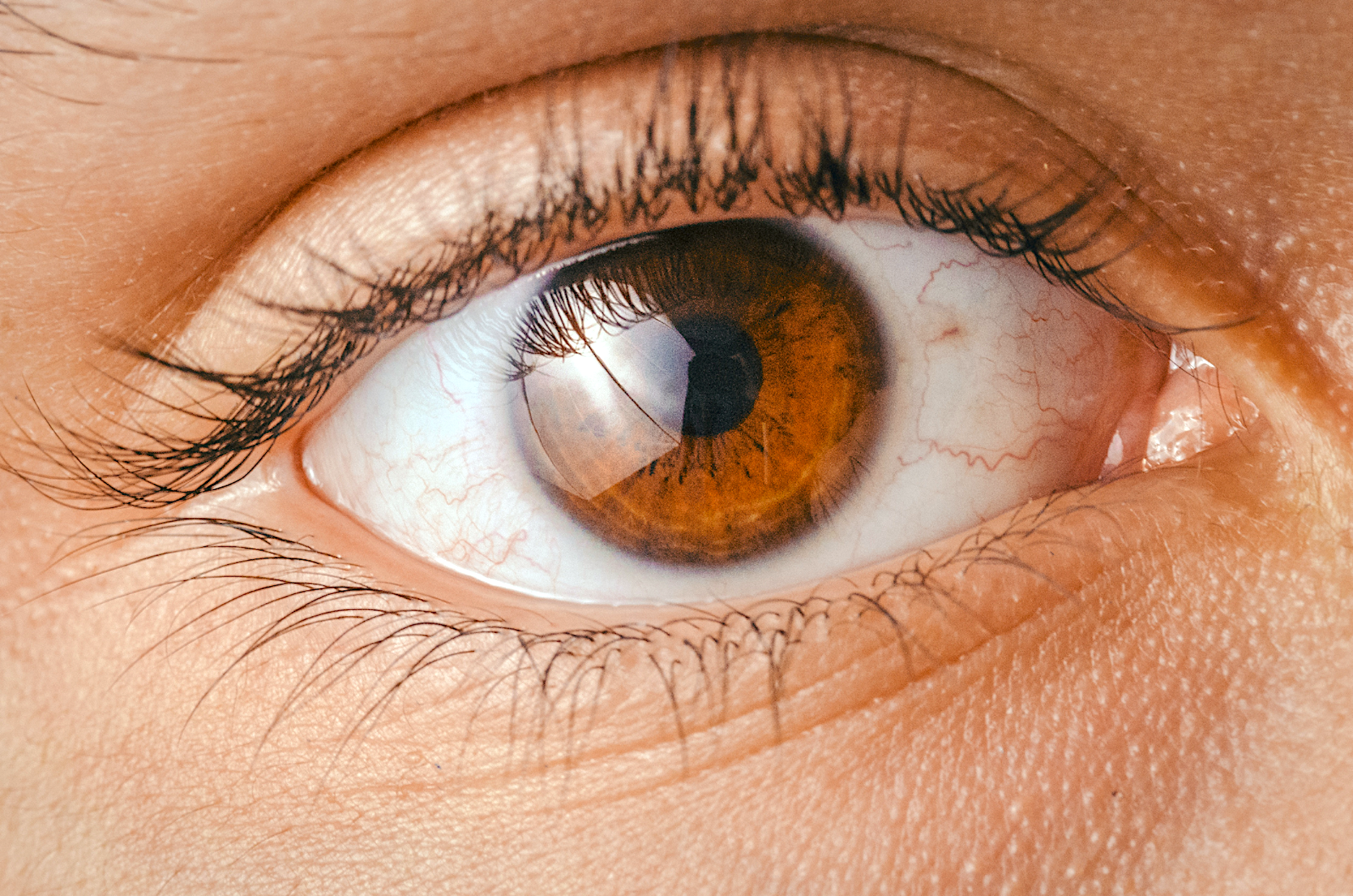
Haematogenic
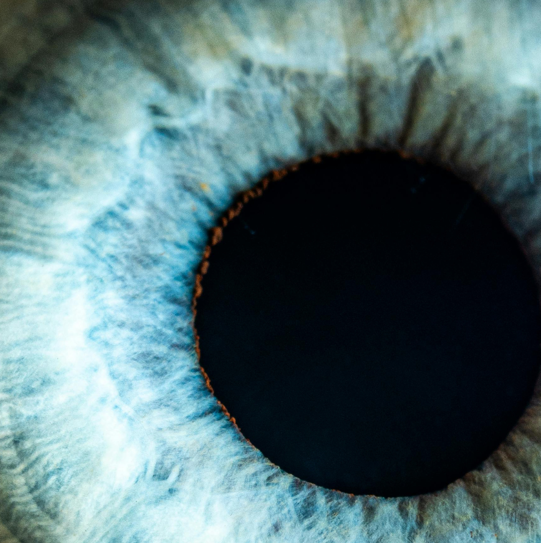
Modern Iridology
Behavioural Iridology
Thanks to the rigorous work of iridology experts, new discoveries are allowing practitioners to better understand markings in their context rather than as simple diagnostic tools.
A
Anterior Border Layer: The iris colour layer. In a blue iris, it is thin (little pigment); in brown eyes, it is thick and pigmented.
Anterior Endothelium: A single layer of microscopic flattened cells. It is a continuation of the posterior surface of the cornea.
Anxiety-Tetanic Structure: Circles and arcs spread throughout the eye, also known as contraction furrows. They also have furrows radiating outward (radial furrows). These rings and grooves are from stress and anxiety patterns from many generations.
Asparagus Lacuna: Usually located in the lower half and indicates a predisposition to chronic degenerative conditions with a tendency toward malignancy. It is topostabile (it is where it is).
B
Biliary or Mixed Constitution: Relates to the gastrointestinal area; radial furrows may be present. Also referred to as the transitory eye.
C
Cardiac Risk Sign (Lacuna): A transversal that runs from the spleen to the heart (it must point towards the heart). The person is more prone to sudden heart problems. It is topolabile (it does not encroach on the heart zone).
Central Heterochromia: Pigmentation concentrated at the collarette in large amounts. This indicates reduced gastric secretions and disturbances of the liver, gallbladder, and pancreas.
Choroid: Long, thin, pigmented tissues (mainly blood vessels) that feed the outer retina.
Cigar Lacuna: May indicate a tumour or growth in the digestive area. It may be in the pancreas or encroaching on the collarette.
Ciliary Body: The portion of the choroid extending into the iris. It is the area of the iris outside the collarette to the iris edge (where you see the iris fibres). It channels nutrients to the iris and produces the aqueous humour.
Closed Lacuna: Closed on both ends; a topostabile genetic marker. If dark and the fibre structure is pulled down, it is more difficult to pull toxins out of a closed lacuna and difficult to get nutrients in.
Conjunctiva: The mucous membrane that lines the eyelids.
Connective Tissue Structure: Loose fibres. A person would have more weakness, such as spinal subluxations or organ prolapses.
Contraction Furrows: Nerve rings – an anxiety tetanic subtype. They are caused by the buckling of the trabeculae in the ciliary zone due to prolonged contraction of the dilator muscle.
Cornea: The transparent continuation of the sclera covering the front of the eye.
Crypt: A diamond-shaped genetic marker indicating long-term, deeply seated toxins. This means degenerative conditions or chronic conditions. Found directly inside or outside of the collarette.
D
Dark Signs: Over-relaxed stroma (exposed stroma layer):
Grey: Slight, not very significant.
Dark Grey: Harder to resolve because of less vital force – can be inherited or acquired through suppression. Indicates toxic residue in the tissues. Waste deposits induce a degenerative process in the organ.
White signs change to dirty white, grey, yellow or brown and become chronic.
Black: The most degenerative and least able to dispel toxins.
Defect of Substance: Small crypts that look like slits or pencil points, found anywhere in the iris. Indicates poor nutrition at the location of the sign.
Density: Equals resistance, which equals structural integrity.
Dilator Muscle: A highly enervated, longitudinal muscle that lies anterior to the posterior epithelium. The sympathetic nervous system moves it to dilate the pupil.
F
Febrile Constitution: A blue-white colour. Indicates hyperactive, oozing conditions, such as skin problems like seborrhoea.
Ferrum Chromatose: Tiger striping indicating liver weaknesses and poor iron absorption.
H
Half Lacuna: When found in the heart area, this shows muscle weakness, affects respiration and affects the heart muscle. Respiration and/or digestion are affecting the heart muscle.
Health Equation: Physical inheritance + environment + spiritual + age = Health.
Haematogenic: A pure brown iris. Indicates an imbalance in blood composition, blood-forming components, and blood circulation.
Honeycomb Lacuna: Webbing or small crypts connected. Most commonly found in the nutritive zone (pupillary zone). If dark inside, it indicates reduced vitality where found. If found in the pancreas, liver, nutritive zone or elsewhere, it creates a good environment for parasites. Perform parasite cleanses starting two days before the full moon.
Hydrogenoid: 'Hydro' means water, so these individuals have drainage problems and are predisposed to allergies. Tophi signify hydrogenoid.
I
Inferior: At the bottom.
Iris Topography of Structures and Functions:
Pupil
Pupillary Border – central nervous system
Pupillary Zone – nutritive zone (stomach, intestines)
Collarette – autonomic nerve wreath
Inner Ciliary Zone or Humoural Zone – fluids, lymph and blood
Mid-Ciliary Zone – major organs and utilisation zone
Outer-Ciliary Zone – bones, skin, external blood and lymph – detoxification and elimination
L
Lacunae: Inherited signs of energy insufficiency that do not necessarily indicate a disease.
Leaf Lacuna: A closed lacuna with veins. Always found in the pancreas, adrenal glands, or gall bladder – the organs of secretion.
Light Signs: Show building, growth, reaction and irritability – indicating good vital force.
Lymphatic Constitution: Mucus afflictions, congested lymph and a predisposition to an overactive immune system.
Lipemic Diathesis or Corneal Arcus:
Arcus senilis, lipemic ring, cholesterol ring
Chemical imbalance
Liver dysfunction; glucose metabolism issues
Mineral deficiencies
M
Medusa or Jellyfish Lacuna: Usually found in the bronchial, lung or kidney areas. Thought to be a cancerous sign and signifies a family history of pathology in the corresponding area. Recommend pap smear, etc.
Medial: Nasal is closest to the nose.
Minor Arterial Circle (Autonomic Nerve Wreath or Collarette): A vascular arterial formation in a ring around the pupillary zone – relates to the intestines and autonomic nerve wreath.
N
Neurogenic Structure: Straight and tight fibres. Indicates physical strength.
Neuronal Netting Lacuna: Resembles a fishnet. Indicates anxiety when found in the lung area. A 'network' of fine fibres below the iris surface. It is topostabile.
O
Open Lacunae: Open on the end. Most often found in the mucous membrane zones, such as the lungs, kidneys, genitalia, bronchus, throat and sinuses. Open lacunae in the heart area indicate a genetic predisposition to heart weakness.
Over Acid Constitution: Light blue is the colour of the eye, and it does not change. Usually indicates problems with an acidic constitution.
P
Perifocal Lightening: Appears as a white band bordering a lacuna, showing long-standing inflammation in the local area.
Pigments of the Iris: Pigments are spots of colour that increase focus on certain areas. They indicate susceptibility to stress and can be topostabile.
Orange: Pancreas, possible liver.
Fluorescent Orange: Gall bladder, pancreas, or liver disturbance.
Straw Yellow: Kidney disturbances.
Dark Brown: Liver.
Pinguecula: A deposit of yellow to clear fatty tissue on the sclera. Indicates disturbed fat metabolism.
Pterygium: Thickened translucent to white tissue growing over the iris, believed to be caused by overexposure to sun, wind, etc. Possible fungal growth.
Pink Radials (or Vascularised Vessels): Associated with great congestion and more weakened conditions in the area.
Polyglandular Structure: Has at least three lacunae attached to the collarette. Reflects hormonal and endocrine gland deficiencies.
Polypose Lacuna: If it pushes into the collarette, it indicates intestinal polyps. It is topostabile to the intestines and indicates sinus problems.
Posterior Epithelium: A darkly pigmented layer of the iris. It prevents light from penetrating through the iris. This black layer can also be seen in crypts, lacunae and fibre rarifications.
Pupillary Border: The small, darker border surrounding the border of the pupil.
Pupillary Sphincter: A muscular band innervated by the parasympathetic nervous system, which causes the pupil to become smaller. It is referred to as the 'stomach ring' when it becomes visible through weak fibres. A white stomach ring indicates acid; a grey ring indicates under-acid.
R
Radial Fibres: Blood vessels coated with a sheath of tissue. Also called trabeculae.
Radial Furrows (Radaii Solaris): Shows a decreased nerve supply to the digestive and intestinal tract, which leaves the corresponding area of the iris vulnerable to toxins.
Reflexives: White stroma fibres that are raised and protrude. They suggest an acute reaction or irritation and indicate that the body has enough energy to release toxins. Possible fevers as the body fights.
Rarefaction: Open fibres or separation of fibres. Indicates decreased vitality in that area and decreased density.
Retina: An extension of the optic nerve and the receptor for visual expressions.
Roof Tile Lacuna: Stair-step appearance; a genetic marker. A 'network' of fine fibres below the iris surface. It is topostabile and indicates a more serious sign.
S
Schnabel Lacuna or Beak Lacuna: Located in the abdominal area and indicates weakness of nerve supply. If it breaks through the collarette or flattens, it is more serious.
Sclera: The white tissue visible as the 'eyeball'. It protects the inner parts of the eyeball.
Scurf Rim Constitution: A dark rim on the outside of the iris. Indicates sluggish skin elimination; the patient needs to sweat.
Sectoral Heterochromia: A section of opposite pigmentation in the iris (for example, a brown sector in a blue iris). It is a genetic marker.
Shading: The formula is: Shading = Reactivity = Vital Force. Shows the organism's ability to react, disperse toxins, and compensate.
Stroma: Iris fibres. A trabecula is an individual fibre within the stroma, and they run radially.
Superior: At the top.
T
Temporal: Closest to the temple.
Tobacco Snuffing: Little dark red-brown dots. Indicate liver issues and are topolabile.
Topolabile: An iris marking that indicates a weakness in a specific organ but can be found anywhere in the iris. Significance is determined by its structure or colour, not by location. For example, a brown pigment indicates liver weakness, even near the heart area.
Topostabile: Markings found in the iris in a specific area of the body which affect that related part of the body. For example, a marking found in the liver area specifically means a weakness in the liver (brown pigment).
Trabecula (plural: Trabeculae): An individual fibre within the stroma layer which runs radially.
Transversals: Appear askew to the normal direction of fibres. They show congestion and a possible sclerotic, a hardening of an organ or tissue, especially due to excessive fibrous tissue growth.
Tulip Lacuna: Usually attached to the collarette, but can be found anywhere. Indicates glandular problems.
U
Uric Acid Diathesis Subtype: Indicates elevated uric acid. Ask who had gout in their family history.
V
Vitreous Humour: The gel between the lens and the retina at the back of the eye. It is approximately 98% water.
W
White Iris Signs: Show overstimulation and hyperactivity. The stroma (vascular) layer has become swollen because of congestion in the blood supply of the organ that is inflamed or congested.
Reference:The Core Curriculum Iris Analysis Manual by Betty S. O'Brian, M.S., N.D.